Neuroscience Crash Course #1
Basic Cell Types: Neurons and Glia
Welcome to the Neuroscience Crash Course, a series of articles that will become the ultimate resource for anyone that wants to learn more about neuroscience and how the brain and body work.
We’ll start here with an introduction to the basic cell types of the nervous system to lay a foundation of knowledge, then dive into how exactly neurons work and the different regions of the brain.
With that out of the way, let’s begin by asking a simple question…
What is a neuron?
Neurons are the simplest unit and building blocks of our nervous system. They're cells that have a unique structure specialized for receiving, conducting, and transmitting electrochemical signals. With almost 100 billion neurons in our bodies, they are much more than just the cells that make up our brain.
They control our muscles, take in sensory information from our environment and relay it to the brain so that we can perceive it, and innervate our various organ systems. Neurons are how the brain communicates with the body and vice versa.
Let’s take a look at the anatomy of a neuron, so we can understand how exactly how they work.
Neurons have four main parts that allow them the unique ability to communicate with each other:
Dendrites
Soma
Axon
Axon Terminals
Like all human cells, neurons contain a nucleus and other organelles needed to maintain life which are stored in the soma (aka cell body). What makes neurons unique though, are the branch-like protrusions coming off of the soma. These are referred to as the dendrites and the axon. Dendrites receive input from other neurons, while the axon carries new messages away from the cell body and delivers it to other neurons.
It is important to keep in mind that neurons typically have many dendrites (most neurons make thousands of synapses), but only one axon. This means they can receive signals from tons of different neurons from all over the brain, though can only pass that signal forward to one specific area.
This path of communication only flows in one direction; in through the dendrites, collects in the soma, passes through the axon, and onto the next neuron. You can think of this internal flow like electricity traveling through a wire, since the internal communication in a neuron is electrical.
How Neurons Communicate
Neurotransmission, or how neurons communicate, involves two mechanisms:
Chemical transmission
Electrical impulses
Chemical transmission is the means by which neurons are able to communicate with each other, while electrical impulses (described above) are how the message is communicated internally.
In response to an electrical impulse one neuron releases small molecules (neurotransmitters), from its axon terminals into the space located between the two, or synapse. These neurotransmitters bind to receptors on the dendrite of the receiving neuron and contribute to the electrical impulse that travels within the neuron, through the body, down the axon, to the terminals.
Different Types of Neurons
Now you know what the “stereotypical” neuron looks like. However, there is so much diversity in their shape and size depending on their function in the body; it can be narrowed down to three main classes that serve different functions within the nervous system.
Sensory Neurons:
These guys do what you’d expect based on the name. They sense things from their environment and send that information to the brain where you are then able to perceive it. They have highly specialized nerve endings that are sensitive to a particular type of stimulus and convert that into electrical impulses.
The image below shows a few examples located in skin. Different nerve endings allow them to sense different inputs, whether it be pressure, stretch, or raw sensation and pain. This class also includes neurons that sense taste, vision, smell, temperature, sound, and even our heads position in space.
In this type of neuron the nerve endings take place of the dendrites, as they’re receiving information externally and not from other neurons.

Interneurons:
These are the primary neurons in the central nervous system. They serve as relay units, and function in the way I described as the “stereotypical neuron” earlier, receiving input from other neurons, and passing that message on to the next.
Their exact function depends on where they’re located in the brain or nervous system generally.
Motor Neurons:
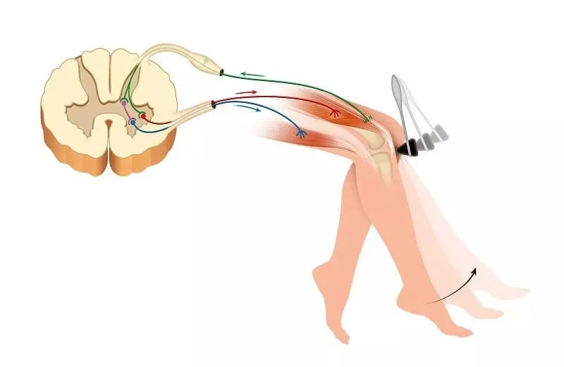
Motor neurons are unique as they elicit a direct effect upon their activation. Instead of sending their signals to other neurons, they activate muscles causing them to contract. Whenever you decide to move, your brain outputs a signal that travels via neurons through the spinal cord and to the effector muscle required to complete that movement.
However, they can also be stimulated by reflexes that bypass the brain entirely. I’ll give you a real life application. Whenever your doctor hits your knee and your leg reflexively kicks out, that signal from your knee bypasses the brain entirely via the spinal cord, causing your quad to contract and hamstring to relax (shown below).

Glia
Glia is a blanket term for the different types of cells that support the function of neurons. They are the unsung heroes of the nervous system. Without them our nervous system would cease to function entirely. In fact, there are more glia than neurons in the brain (there are trillions of glia), and there are five main types:
Astrocytes
Microglia
Ependymal cells
Oligodendrocytes
Schwann cells
Let's go over the function of each.
Astrocytes are the most abundant type of glia, and they have a wide range of function in the brain. This includes, regulating the blood-brain barrier (BBB), providing structural support for neurons, synthesizing neurotransmitters, reuptake of extracellular neurotransmitters, forming scar tissue after injury in the central nervous system (CNS), and more.
Note - The CNS includes the brain and spinal cord, while the peripheral nervous system (PNS) includes all other neurons not present in the brain or spinal cord.
These cells serve as the primary support cells for neurons, as well as have the important job of maintaining the BBB, the first line of defense for what gets to enter the brain. Astrocyte's end feet wrap around blood vessels to help maintain their tight junctions (shown below). The integrity of these junctions limit what can and can't enter the brain, keeping out toxins, pathogens, and other harmful substances.

Microglia are the immune cells of the brain. They react to pathogens and damage. When a threat is detected, they rapidly change shape (or activate) and move to the area of need.
These cells will 'eat' dead cells, debris or pathogens via phagocytosis to clean house in the brain.
Ependymal cells form the lining of ventricles, a series of connected cavities filled with cerebrospinal fluid (CSF) in the brain and spinal cord. The main function of these glia is to produce and secrete CSF into ventricles, as well as assisting in its circulation using the cilia present on their exterior.

The last 2 have very similar functions as they both myelinate the axons of neurons, but they operate in slightly different ways (and locations).
Oligodendrocytes myelinate neurons in the CNS. They can myelinate many parts of several different axons, which is their biggest difference from the next type. They also contribute to maintenance and repair of axons in the CNS.
Schwann cells myelinate one part of a single axon in the peripheral nervous system (PNS), so it takes multiple Schwann cells to cover one whole axon. They also maintain and regenerate the axons in the PNS is damage occurs.
Myelin is the white fatty substance that surrounds the axons of most neurons, allowing the action potential to travel faster. This is why the axon tracts are known as 'white matter' in brain tissue.

Note - Not all neurons have myelinated axons, in particular some sensory neurons in the PNS don't, but all motor neurons do since we want that message traveling as fast as possible.
Proper neuronal function requires neurons and glia to work together. They all exist in the same micro-ecosystem that is our nervous system, and neurons can't function to the best of their ability (or at all) without glia there to support and protect them.
Conclusion
With that, we have an introduction to the primary cell types that make up the human nervous system, neurons and glia. This content barely scratches the surface of neuroscience, but it’s important to have a know these things before we dive deeper into neuronal function and other topics.
A brief recap:
Neurons are the primary cells that make up the nervous system
They communicate with each other using a combination of electrical and chemical signals
There are three main types of neurons
Sensory Neurons
Interneurons
Motor Neurons
There are 5 main types of glia that work to support neurons
Looking Forward
With this base of knowledge we can now begin to dig into the specifics of how neurons work. The next installments will cover:
Membrane Potential and the Action Potential
Neurotransmitters and Synaptic Transmission
Different brain regions and their function
After that, you should have a much greater understanding of the nervous system and how exactly neurons work.
Moving forward, all “Neuroscience Crash Course” articles will be for paid subscribers only. A subscription is only $5 a month ($50 for the year) and I believe it is more than reasonable for the amount of content you’ll get in return.
Along with the continuation of this series, I have other articles planned aimed at helping you optimize neurotransmitters and brain health/function using diet, lifestyle, and obviously supplements/nootropics.
There is still free content coming as well, so make sure to subscribe to not miss any new articles. Reminder that if you sign up as a free or paid sub you’ll get my “Intro to Neurotransmitters” eBook sent to you immediately, which includes an in depth intro to all the main neurotransmitters in the brain.
That’s all for now. Thank you for reading.
Link to Neuroscience Crash Course #2


